Bring Intelligence to your Clinical Workflows & Patient Management
Leverage Discovery® for Clinics powered by CE-marked & research use only AI models to enhance patient treatments & deliver the right treatment, at the point of care.
.png)


Discovery® for Clinics is made for Ophthalmologists & Optometrists
See how it transforms clinical workflows

Effortlessly Manage All Patient Data
Tired of using multiple viewers to access your patient data? View and navigate all your data, including OCTs, Fundus images, and more, in a single platform enabling data harmonisation & interoperability.
.png)
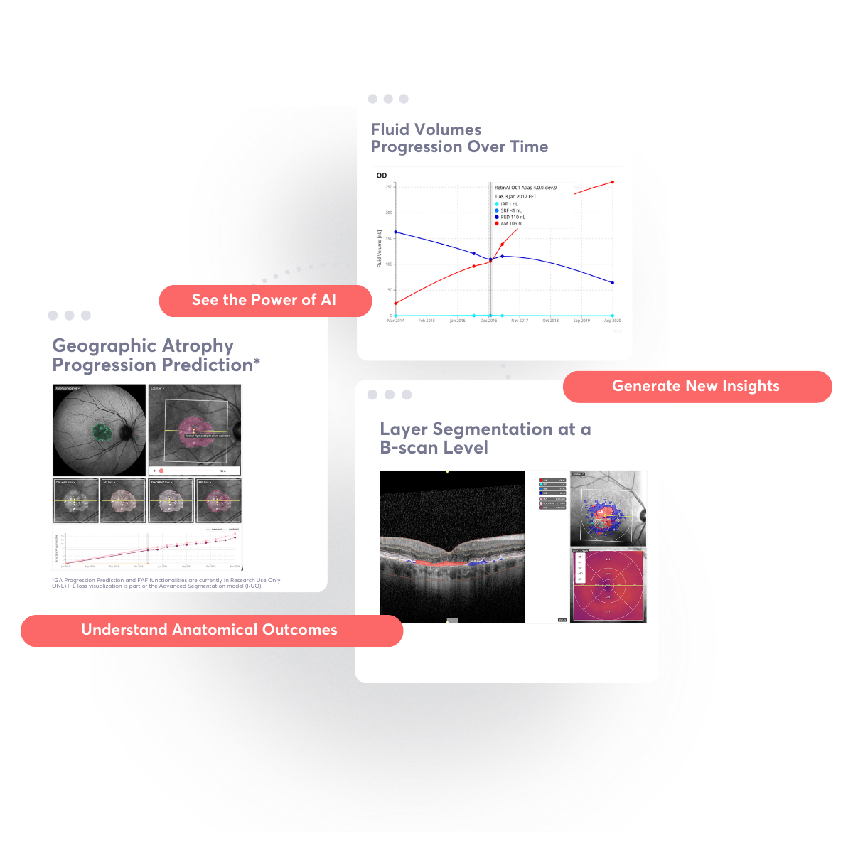
Transform Disease Evaluations with AI
Harness AI for enhanced insight into disease progression and prediction, precise retinal layer segmentation, and accurate fluid volume assessment.
.png)
Elevate Tele-ophthalmology Experience
Ensure secure, real-time peer referrals on a unified platform, enhancing communication and patient care without the hassle.
Choose the package tailored for you
Redefine your Clinical Workflows & Patient Management
Unlock data insights from easy to use Artificial Intelligence to optimize outcomes, treatments & general costs.
Access & Navigate your Patient Data in a Single Platform
Discovery enables you to have a full overview on the patient information available: view all OCTs, Fundus images, DICOM datasets, PDFs, JPEG, PNG & TIFF files in one single place - no need for multiple viewers.

Transform your Disease Evaluations with our CE-marked AI Models
Unleash the power of AI to analyze OCT volumes, annotate images, craft eCRFs, and gather essential insights for future reference with our Fluid & Layer Segmentation AI models, our Advanced Semgentation model and GA progression prediction model.
Upload your Clinic Data and Let the Magic Begins
Once data from your clinic is uploaded, it is automatically harmonised & structured at scale and processed in real time with AI for endpoints & measures, and biomarkers identification.
.png)
Experience a Turnkey Tele-Ophthalmology Platform
Unlock swift, secure data sharing and immediate evaluation on a unified platform for real-time peer referrals. No more non-secured emails, your data will be securely stored and shared with easy to give access.
Provide Your Patients With the Ability to Own Their Clinical Data
With the "Passport" feature, your patients can access their clinical data easily through Discovery, and you can improve patient engagement by creating remote questionnaires.

Enhance Referral Decisions & Patient Management
Get accessible and easy to use AI for better referrals, better patient engagement and new revenue streams.
Access & Navigate your Patient Data in a Single Platform
Discovery enables you to have a full overview on the patient information available: view all OCTs, Fundus images, DICOM datasets, PDFs, JPEG, PNG & TIFF files in one single place - no need for multiple viewers.
Transform your Disease Evaluations with our CE-marked AI Models
Unleash the power of AI to analyze OCT volumes, annotate images, craft eCRFs, and gather essential insights for future reference with our Fluid & layer segmentation models.
Upload your Clinic Data and Let the Magic Begins
Once data from your clinic is uploaded, it is automatically harmonised & structured at scale and processed in real time with AI for endpoints & measures, and biomarkers identification.
Experience a Turnkey Tele-Ophthalmology Platform
Unlock swift, secure data sharing and immediate evaluation on a unified platform for real-time peer referrals. No more non-secured emails, your data will be securely stored and shared with easy to give access.
Provide Your Patients With the Ability to Own Their Clinical Data
With the "Passport" feature, your patients can access their clinical data easily through Discovery, and you can improve patient engagement by creating remote questionnaires.
An online platform powered by Certified & RUO AI models
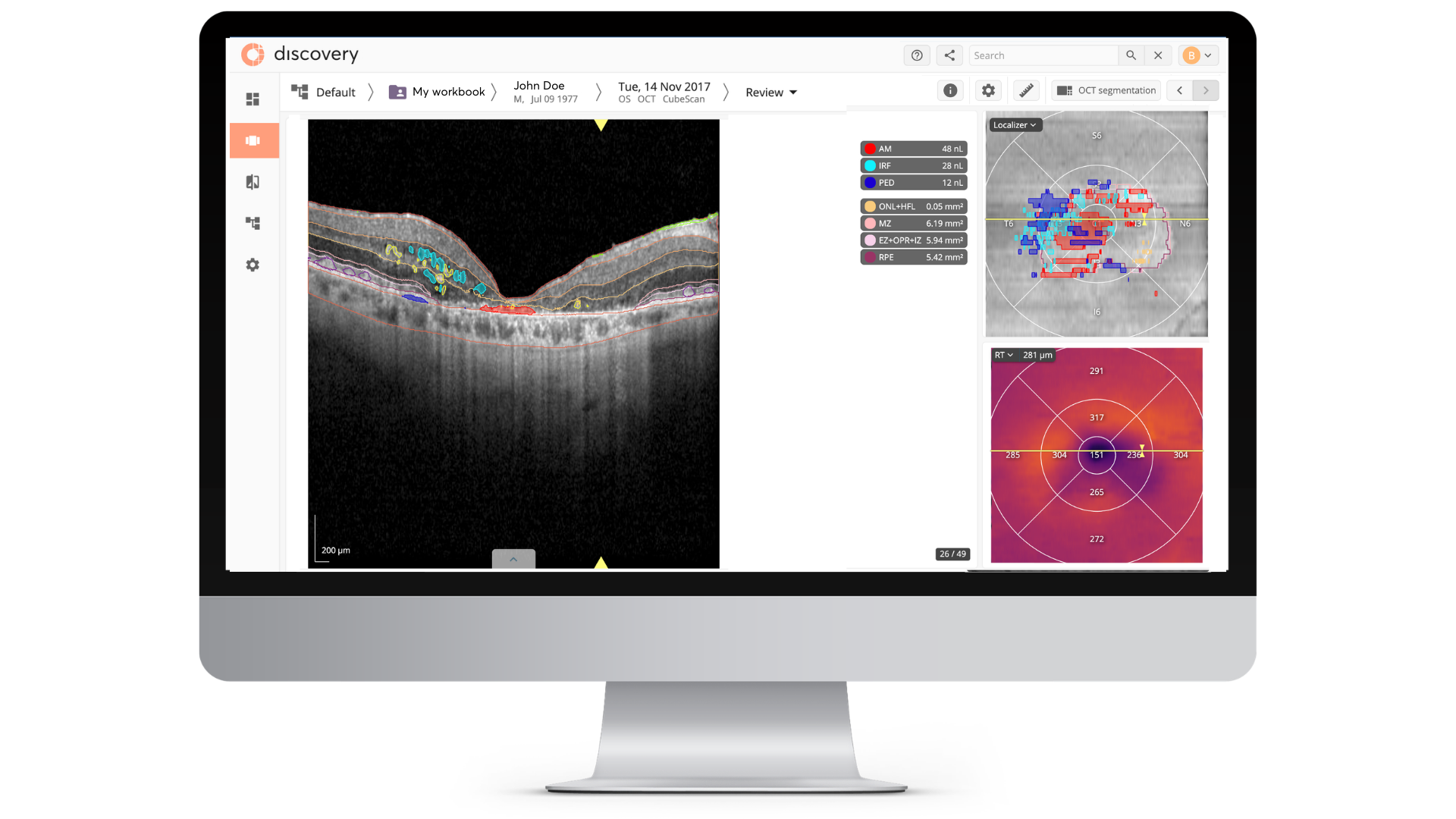
NEW: RetinAI OCT Atlas®
Harness our CE-marked AI Model for precise Layer Segmentation & Fluid Segmentation and Quantification of fluid volumes, including Intraretinal Fluid (IRF), Subretinal Fluid (SRF) and Pigment Epithelium Detachment (PED)/DRUSEN and Amorphous Material, including hyperreflective material in the subretinal space (e.g., fibrosis).
.png)
AI-Powered & Precise Layer Segmentation
RetinAI OCT Atlas® provides measurements for the following layers:
• Retinal Nerve Fiber Layer (RNFL);
• Ganglion Cell Layer (GCL) + Inner Plexiform Layer (IPL);
• Inner Nuclear Layer (INL) + Outer Plexiform Layer (OPL);
• Outer Nuclear Layer + Henle’s Fibre Layer (ONL+HFL)
• Myoid Zone (MZ)
• Photoreceptors: Ellipsoid Zone (EZ) + Outer Photoreceptor Segment (OPR) + Interdigitation Zone (IZ) (EZ+OPR+IZ)
• Retinal Pigment Epithelium (RPE)
• Choriocapillaris and Choroidal Stroma (CC+CS)
.png)
Enface view provides thickness measurements and visually highlights attenuation or thinning of layers
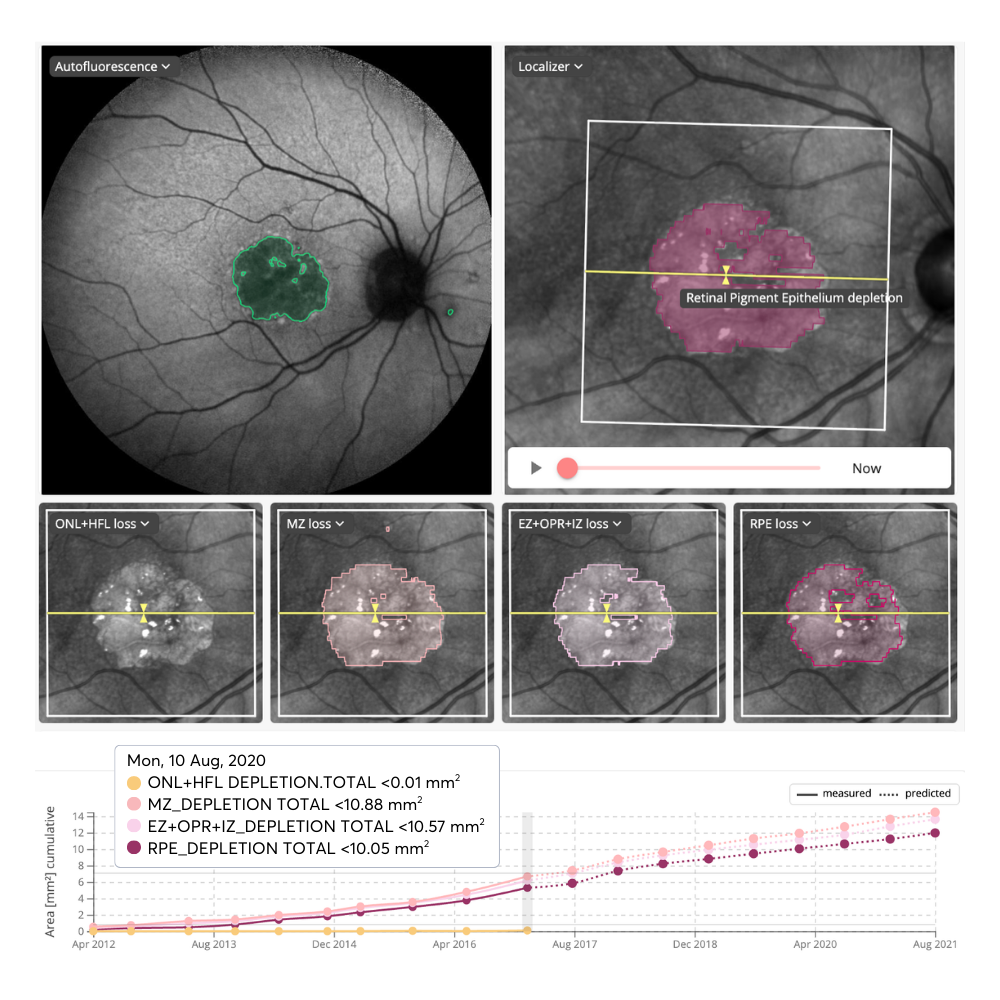
Advanced OCT Segmentation (RUO)
This AI Model module significantly expands analytical capabilities by adding automated detection and quantification for HRF, SDD, and ERM. This Research Use Only suite provides comprehensive thickness and volume measurements for these additional biomarkers, alongside specialized ONL+HFL, Photoreceptor & RPE depletion maps
and advanced post-processing tools like automated fovea localization. It is designed for efficient scalability, in-depth insights necessary for high-level clinical research and longitudinal disease progression studies.
and advanced post-processing tools like automated fovea localization. It is designed for efficient scalability, in-depth insights necessary for high-level clinical research and longitudinal disease progression studies.
Macula Biomarkers AI Model
This model provides probablility of presence/absence of retinal biomarkers at a B-scan level in volumes. Biomarkers identified are: Fluids (SRF, IRF and Fibrous PED), Hyper reflective Foci (HF), Drusen, Reticular Pseudodrusen (RPD), Epiretinal Membrane (EPM), Geographic Atrophy (GA) and Outer Retinal Atrophy (ORA).

Geographic Atrophy Progression RUO*
This model provides segmentation of GA from OCT scans and simulation of atrophy progression based on visit data.
Predict personalized retinal layer depletion with a single OCT scan. This model simulates the progression of atrophy and EZ, MZ, and RPE layer depletion over 54 months (in 6-month increments) for patients with GA secondary to AMD.
Automated Outputs:
• Progression Maps: En-face depletion areas for EZ, MZ, and RPE at 9 time points spaced every 6 months (mm2).
• ETDRS Analytics: Area measurements (mm2) per ETDRS segment across all 9 time points spaced every 6 months (mm2).
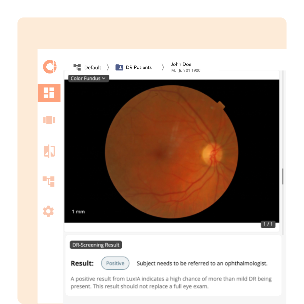
Diabetic Retinopathy Screening with LuxIA®
AI-Powered mtmDR Screening LuxIA® is a CE-marked, Class IIb AI medical device designed to screen adult diabetic patients for more-than-mild diabetic retinopathy (mtmDR). By processing 45° color fundus images (Topcon or equivalent), the model identifies patients likely to have mtmDR with 97% sensitivity and 95% specificity, supporting timely referrals to specialists. LuxIA runs seamlessly within the RetinAI Discovery® platform to enable earlier detection and help prevent vision loss.