RetinAI OCT Atlas® is now CE-marked for Clinical Use: one algorithm for AMD, DR, DME, and Glaucoma
RetinAI’s most comprehensive OCT analysis algorithm is now certified as a medical device under EU MDR (2017/745), covering four retinal disease areas, validated across the major OCT device makers, and available for routine clinical use in the EU.
Bern, Switzerland – 24 June, 2026 – RetinAI (Ikerian AG) announced the launch of RetinAI OCT Atlas®, its most comprehensive OCT analysis algorithm, certified as a Medical Device Software under the EU Medical Device Regulation (MDR 2017/745) for clinical use.
This certification follows the completion of the DILL study, a multi-device, multi-grader clinical validation study involving 124 patients, three OCT devices (Heidelberg Spectralis, Topcon 3D OCT-2000, Topcon Triton), and ten expert graders assigning three independent annotations per scan. Every pre-specified performance and safety endpoint was met. The model's segmentation accuracy was in line with inter-grader agreement (meaning it quantifies retinal biomarkers as reliably as expert graders measuring against each other) and does so in <1 second per full OCT cube scan, against 1.2 hours for the fastest human grader.
RetinAI OCT Atlas® is available now as part of the RetinAI Discovery® platform. In the EU, its CE mark under MDR 2017/745 makes it available for both routine clinical use and research. In the U.S., it is available for Research Use Only (RUO).
What RetinAI OCT Atlas® measures
RetinAI OCT Atlas® automatically segments and quantifies 12 retinal features including retinal layers, fluids, and biomarkers, from the nerve fibre layer to the choroid, including fluid types (IRF, SRF, PED), drusen and amorphous material. It tracks changes across visits without manual input, surfacing progression in structures like the Ellipsoid Zone, Myoid Zone, and RPE that predict disease activity before vision loss occurs.
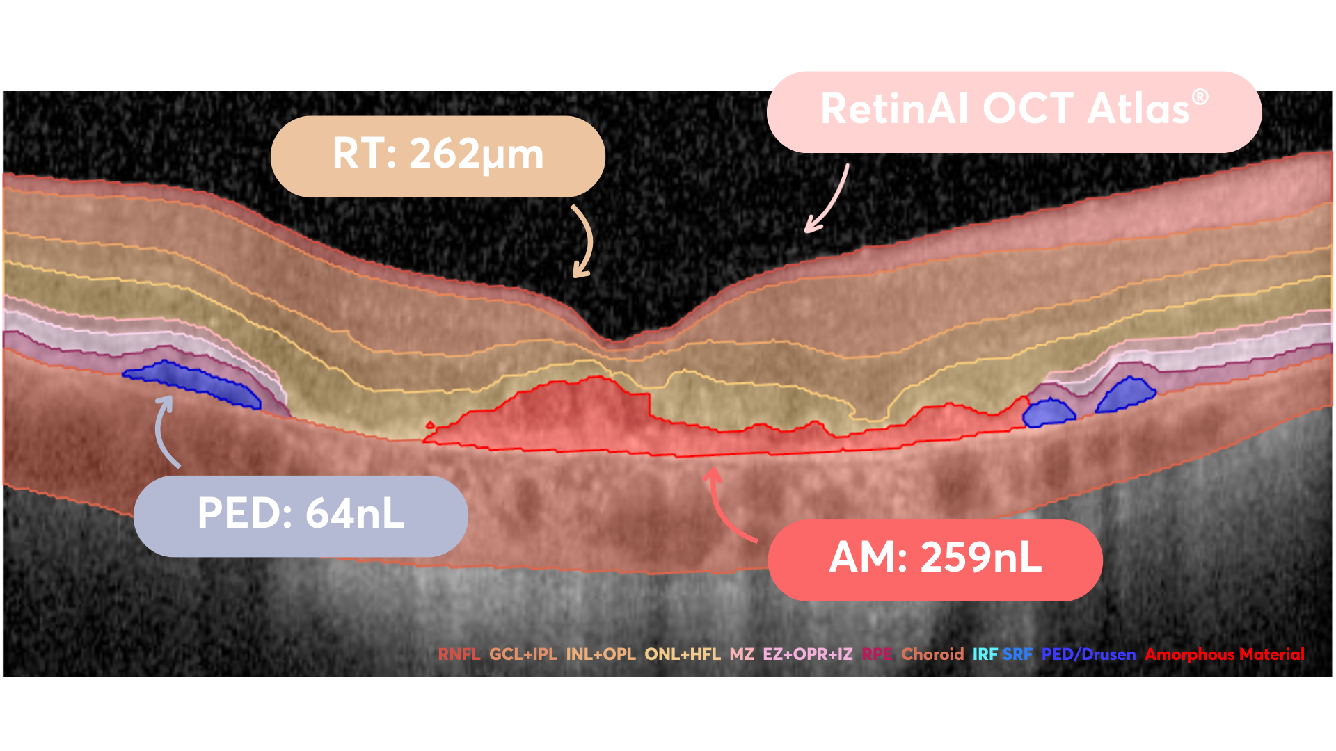
Approved indications: AMD, DR, DME, and Glaucoma
RetinAI OCT Atlas® is approved within RetinAI Discovery® for the following indications: iAMD, AMD, neovascular AMD (nAMD), Geographic Atrophy (GA), Diabetic Retinopathy (DR), Diabetic Macular Edema (DME), and Glaucoma, the newest addition to the certified portfolio.
Glaucoma represents a particularly time-sensitive indication. RNFL thinning and ganglion cell layer (GCL+IPL) loss correlate directly with visual field deterioration. Glaucoma is frequently asymptomatic until late stage, and the structural damage is permanent once it occurs. Consistent, quantitative monitoring of RNFL and GCL+IPL must begin before symptoms appear, which requires repeatable, objective measurement across visits, not qualitative review.
Tracking disease at the structure level
In conditions like Geographic Atrophy, the outer retinal layers don't fail together, the Myoid Zone, Ellipsoid Zone, and RPE degrade at different rates and at different stages. RetinAI OCT Atlas® tracks each structure separately, so changes that matter clinically aren't averaged away. Area loss ratios can be calculated to understand progression rates.
“The launch of RetinAI OCT Atlas® is a significant step forward in how we quantify retinal pathology. By decoupling complex biomarkers and offering specialized analysis for outer retinal layers like the Myoid Zone, Ellipsoid Zone and Retinal Pigment Epithelium, we are providing clinicians with the granular data necessary to understand disease activity at a much more precise level. Our goal has always been to turn complex imaging into clear, actionable insights, and this unified model achieves exactly that,”
said Dr. Sandro De Zanet, PhD, Co-Founder and Chief Scientific Officer at RetinAI / Ikerian AG.
Integration with RetinAI Discovery®
RetinAI OCT Atlas® is part of the RetinAI Discovery® platform. OCT scans from Heidelberg, Topcon, and Zeiss devices are automatically pulled in via Discovery Connect®, anonymized, and analyzed, no manual import required. Progression tracking updates as new scans arrive.
For research workflows requiring additional biomarkers (Subretinal Drusenoid Deposits, Hyperreflective Foci), these are available through a separate Advanced Segmentation AI Model, designated Research Use Only (RUO) in the EU and US, not for clinical decision support.
The RetinAI Discovery® platform is FDA 510(k) cleared in the United States. RetinAI OCT Atlas® and all RetinAI AI algorithms are designated Research Use Only (RUO) in the US, they are not FDA cleared for clinical decision support.
About Ikerian AG and RetinAI Inc. U.S.
Established in 2017, Ikerian AG (formerly RetinAI Medical AG) and its subsidiary, RetinAI U.S. Inc. (‘Ikerian’, ‘RetinAI’ and, together, ‘the company’), develops software solutions to accelerate clinical, research and pharmaceutical workflows globally using advanced machine learning and computer vision. Focused on the ophthalmology and optometry market, RetinAI builds tools to collect, organize and analyze health data from the eyes, enabling healthcare professionals to make the right decisions sooner in healthcare. RetinAI's international team leverages its clinical, technical, and scientific expertise to foster the transition from reactive to preventive medicine for severe eye diseases. A commercial stage company with growing sales from RetinAI Discovery®, RetinAI has established collaborations and partnerships with leading ophthalmology clinical networks and pharmaceutical companies. About Ikerian AG www.ikerian.com and RetinAI Inc. US www.retinai.com.